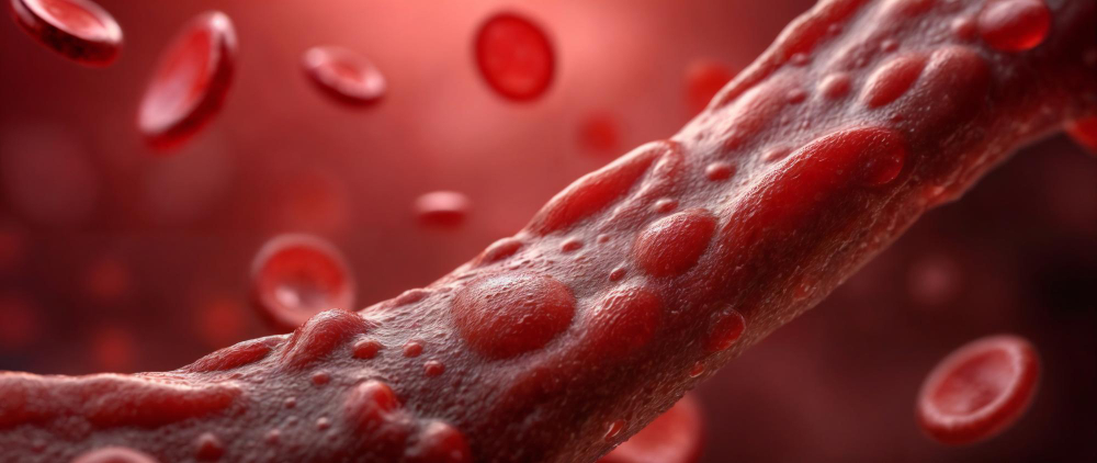
Renovascular conditions narrow the arteries that carry blood to the kidneys, often causing high blood pressure or kidney damage. Diagnosis relies on imaging tests such as duplex ultrasound, CT angiography, MR angiography, and catheter angiography. Treatment options for renovascular conditions include medication, lifestyle changes, angioplasty with or without stenting, and surgery.
A renovascular condition can develop quietly for years before it makes itself known. By the time symptoms appear — usually as difficult-to-control high blood pressure or declining kidney function — the underlying cause may already be well advanced. Understanding how these conditions are identified and what can be done about them gives you the knowledge to act early and ask the right questions at your next appointment.
This guide walks through the diagnosis of renovascular conditions and the treatment options currently available, drawing on guidance from the National Institutes of Health and the Mayo Clinic.
Diagnosis of Renovascular Conditions
Pinpointing a renovascular condition takes more than a blood pressure reading. Because the renal arteries sit deep in the body, your doctor will typically begin with a physical exam and blood and urine tests to assess kidney function, then move on to imaging to get a clearer picture. A vascular specialist will often lead or advise on this process, particularly when initial tests suggest a narrowing in one or both renal arteries.
Four main imaging tests are used to diagnose renovascular conditions:
Renal Artery Duplex Ultrasound
Duplex ultrasound combines standard ultrasound with Doppler technology, which measures the speed and flow of blood through the renal arteries. It is non-invasive, does not require radiation, and carries a low cost compared to other imaging methods. The test can reveal blockages and detect blood moving more slowly than it should — a sign that a narrowing may be restricting flow. Duplex ultrasound is one of the most commonly used first-line imaging tests for renal artery stenosis.
CT Angiography
Computerized tomographic angiography, or CTA, uses a combination of X-rays and computer processing to generate detailed cross-sectional images of the renal arteries. A contrast dye is injected into a vein in your arm to make the arteries more visible. CTA is less invasive than catheter-based procedures and produces results quickly. However, the contrast medium used can be problematic for patients who already have reduced kidney function, so your doctor will weigh this carefully before recommending it.
MR Angiography
Magnetic resonance angiography (MRA) produces detailed images using radio waves and magnetic fields — no X-ray radiation involved. In addition to showing the structure of the arteries, MRA can provide a functional assessment of blood flow and organ activity. Like CTA, MRA may use a contrast agent, though this is generally avoided in patients with poor kidney function due to the risk of complications.
Catheter Angiography
Catheter angiography is widely regarded as the gold standard for diagnosing renal artery stenosis. A thin, flexible tube is guided through the arteries — typically from the groin — to the renal artery, where a contrast dye is injected, and X-ray images are taken. The procedure is more invasive than the others and carries a small risk of bleeding or arterial injury. Its significant advantage is that treatment can be delivered during the same procedure if a significant blockage is found.
Treatment Options for Renovascular Conditions
Once a diagnosis has been confirmed, the goal of treatment is to prevent further narrowing, manage blood pressure, and protect kidney function. Critical renal artery stenosis is a reduction of more than 60 percent in the diameter of the artery — the threshold at which active treatment is typically recommended. A qualified vascular specialist will determine the best approach based on the severity of the condition and the patient’s overall health.
Medical Management
For many patients, treatment begins with medication and lifestyle adjustments. Two types of blood pressure medication — ACE inhibitors and angiotensin receptor blockers (ARBs) — have shown effectiveness in slowing kidney disease progression. Diuretics, beta blockers, and calcium channel blockers may also be part of the regimen. Alongside medication, cholesterol-lowering drugs and low-dose aspirin are often prescribed to manage underlying atherosclerosis.
Lifestyle changes support these efforts. Quitting smoking, reducing sodium intake, maintaining a healthy weight, and staying physically active all help slow the buildup of plaque in the arteries. These changes do not reverse existing damage, but they can meaningfully slow its progression.
Percutaneous Transluminal Renal Angioplasty (PTRA) With or Without Stent
When medication alone is not enough, a minimally invasive procedure called percutaneous transluminal renal angioplasty (PTRA) may be recommended. A catheter with a small balloon at its tip is guided to the narrowed section of the renal artery and inflated to widen it. In many cases, a small mesh tube called a stent is then placed inside the artery to hold it open and maintain blood flow.
The approach varies depending on the underlying cause. Patients with renal artery stenosis caused by fibromuscular dysplasia (FMD) may respond well to angioplasty alone, without a stent. For those whose stenosis is caused by atherosclerosis, angioplasty with stenting tends to produce better outcomes.
Surgical Options
Surgery is less commonly used today but remains an option when other treatments have not produced results, or when the anatomy of the artery makes a catheter-based approach impractical. Two main surgical procedures exist:
- Endarterectomy: The plaque is removed directly from inside the artery, leaving the inner wall smooth and clear.
- Bypass surgery: A vein or synthetic graft is used to create an alternative blood flow route around the blocked artery, connecting the kidney to the aorta.
Both procedures carry a higher risk than angioplasty and require full anesthesia, so they are typically reserved for more complex cases.
Taking the Next Step
Renovascular conditions are manageable. The earlier they are identified and treated, the better the chances of preserving kidney function and keeping blood pressure under control. You do not have to navigate this process alone.
If you or a loved one has been diagnosed with a renovascular condition — or if high blood pressure has been difficult to control despite multiple medications — speaking with a vascular specialist is a meaningful next step. Our Vascular Health team is here to connect you with the right care in your area. Whether you are searching for a vascular clinic in Brooklyn or need guidance on what to expect from your first consultation, we can help you find qualified, compassionate support close to home. Reach out today by calling 1-347-384-5690 or visiting us at 1797 Pitkin Avenue, Brooklyn, New York 11212 to get started.