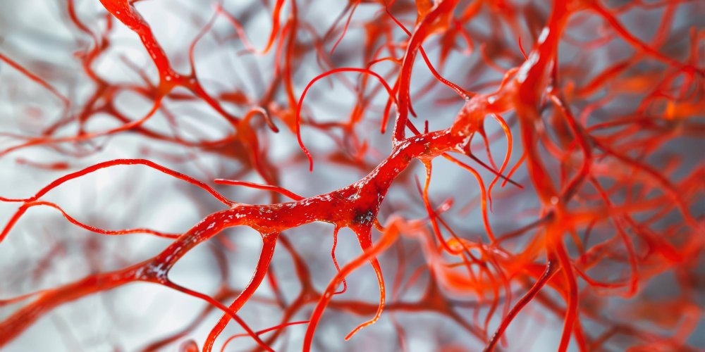
Arteriovenous malformation (AVM) is a congenital condition in which abnormal connections form between arteries and veins, disrupting normal blood flow and raising the risk of bleeding in the brain or spinal cord. Key ways to manage congenital risks of arteriovenous malformation include regular monitoring, medication, and surgical or minimally invasive treatment guided by a vascular surgeon.
A diagnosis of arteriovenous malformation can feel overwhelming. You may have discovered it after a seizure, a severe headache, or a neurological episode — or it may have appeared on an imaging scan before any symptoms emerged at all. Either way, the concern is real, and it deserves a clear, honest answer. Arteriovenous malformation is a congenital condition, meaning it is present from birth, and understanding how to manage its risks is one of the most important steps you can take for your long-term health.
What Is Arteriovenous Malformation?
Arteriovenous malformation occurs when arteries and veins form abnormal connections, bypassing the capillaries that normally slow and regulate blood flow. Without that regulation, blood moves at high pressure directly from arteries into veins. Over time, that pressure can weaken vessel walls and raise the risk of rupture or bleeding.
AVMs most commonly affect the brain and spinal cord, though they can develop elsewhere in the body. Many people live with an unruptured AVM for years without symptoms. Others experience headaches, seizures, muscle weakness, or neurological changes. The unpredictability of this condition is part of what makes early evaluation and ongoing management so important.
How Are Congenital Risks of Arteriovenous Malformation Assessed?
Before any treatment decisions are made, your care team needs a clear picture of the AVM — its size, location, and how it behaves under normal blood flow conditions. A vascular surgeon or neurovascular specialist typically uses one or more of the following imaging tools:
- MRI and MRA (Magnetic Resonance Angiography): These provide detailed images of blood vessels and surrounding brain tissue without radiation exposure.
- CT Angiography: A fast, effective tool for identifying bleeding or structural abnormalities.
- Cerebral Angiography: Considered the gold standard for AVM evaluation, this procedure maps blood vessels with precision and helps guide treatment planning.
The findings from these tests help determine whether the AVM poses an immediate risk — and which approach to managing congenital risks of arteriovenous malformation is most appropriate for your situation.
What Are the Main Ways to Manage Congenital Risks of Arteriovenous Malformation?
There is no single path that works for every person. Treatment decisions depend on the AVM’s size, location, your overall health, and whether symptoms are present. A qualified vascular surgeon or neurovascular specialist will review all of these factors before recommending a course of action.
Observation and Monitoring
For small, asymptomatic AVMs in areas difficult to treat surgically, careful observation may be the most appropriate approach. This involves regular imaging to track any changes in the AVM’s size or behavior over time. It is not a passive choice — it is a deliberate, evidence-based decision that keeps you informed and prepared.
Medication Management
While medication cannot eliminate an AVM, it plays an important role in managing symptoms. Anti-seizure medications help reduce seizure activity. Pain management strategies address chronic headaches. Your care team will tailor these to your needs and adjust them as your condition evolves.
Surgical Removal
When an AVM is accessible and the surgical risk is acceptable, direct removal offers the possibility of a permanent cure. A neurosurgeon excises the abnormal vessels while working to preserve surrounding healthy tissue. Recovery varies depending on the AVM’s location and the individual patient’s baseline health.
Stereotactic Radiosurgery
Radiosurgery — including treatments such as Gamma Knife or CyberKnife — delivers focused radiation to the AVM, gradually causing the abnormal vessels to close off over a period of two to three years. It is a non-invasive option well suited to small, deep AVMs that carry high surgical risk.
Endovascular Embolization
This minimally invasive procedure involves threading a catheter through the blood vessels to inject a material that blocks blood flow into the AVM. Embolization is often used alongside surgery or radiosurgery to reduce the AVM’s size and lower procedural risk, rather than as a standalone cure.
Taking the Next Step With Your Care Team
Living with arteriovenous malformation does not have to mean living in uncertainty. With the right evaluation, a clear treatment plan, and consistent follow-up, most people with AVM can manage their condition and protect their quality of life.
If you are in the New York area, connecting with our vascular team give access to the diagnostic tools your situation requires without unnecessary delays. We work with patients and their families through every stage of evaluation, treatment, and recovery. You do not have to face this alone. Reach out today by calling 1-347-384-5690 or visiting us at 1797 Pitkin Avenue, Brooklyn, New York 11212 to speak with a vascular surgeon who can guide you forward with clarity and care.