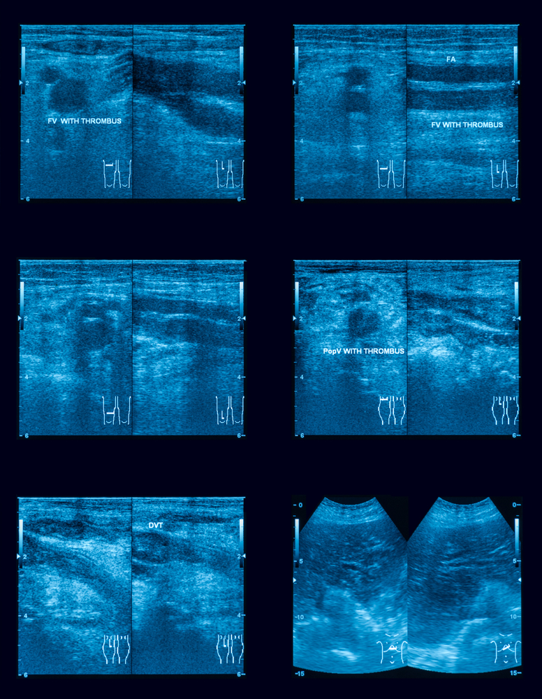
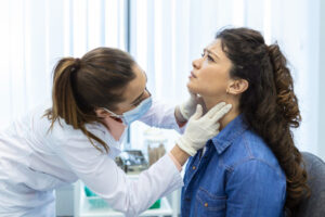
Venous Vascular Ultrasound is a widely used, noninvasive imaging technique that helps detect problems in blood flow through your veins, especially in the legs. But how does it differ from venography, another method for assessing vein health? Both procedures serve as valuable diagnostic tools for identifying conditions like deep vein thrombosis (DVT), chronic venous insufficiency, or varicose veins—but they differ significantly in how they are performed, their risks, and their comfort level for patients.
Venous Vascular Ultrasound uses sound waves to create real-time images of the blood flowing through your veins. It’s a safe and painless procedure that doesn’t involve radiation or the injection of contrast dye, making it a preferred first-line choice for many patients. In contrast, venography is a more invasive technique involving X-rays and injected contrast dye, which may not be suitable for everyone—especially those with kidney problems or allergies to iodine-based contrast.
Choosing between Venous Vascular Ultrasound and venography often depends on the specific condition being investigated, your medical history, and your physician’s recommendation. Some cases may require a more detailed look that only venography can provide, while others can be effectively managed with a simple, fast ultrasound.
If you’re experiencing leg pain, swelling, or other symptoms of poor venous circulation, it’s important to undergo the right imaging study for accurate diagnosis and treatment. Venous Vascular Ultrasound may be the first step in identifying underlying vascular issues and preventing more serious complications.
Book your consultation today with the best cardiologists in Brooklyn and get personalized advice on which imaging method is best for your vascular health.
Venous Vascular Ultrasound vs Venography
Both these procedures are used to diagnose conditions that affect your blood flow in the veins including blood clots; especially, in the veins of the arm, leg, and pelvis. However, venous vascular ultrasound uses sound waves and venography uses X-ray imaging for diagnosis.
Healthcare providers often prefer vascular ultrasound to diagnose blood clots in the veins. However, venography is recommended when ultrasound can’t provide much information. Venography is also used to plan and guide venous disease treatments.
While ultrasound doesn’t use any ionizing radiation and causes no harmful effects, venography is invasive and uses radiation. The provider uses a small needle to puncture your skin to access your veins and inject a contrast dye into your veins to make the blood flow visible on the X-ray. In the X-ray, a small dose of radiation is used. Most people may experience mild discomfort during the test, but it has no serious side effects. But it is not safe for some people such as pregnant women. So, talk with your provider before having a venography and they decide whether this test is appropriate for you or not.
Uses
Both have similar uses; however, venography is recommended only when your venous ultrasound provides less information than is needed to make a diagnosis. The uses are:
- Used to diagnose conditions such as deep vein thrombosis, which is caused by blood clots, especially in the legs which cause swelling and pain. Additionally, varicose veins are also identified with both tests that can damage valves and cause abnormal blood flow.
- It is also used to diagnose blood flow problems in your pelvic area which include pelvic congestion syndrome, nutcracker syndrome, and varicoceles.
- It is used to check flow and real-time imaging during treatments and surgeries such as thrombolytic therapy, thrombectomy, and varicose vein stripping and ligation. However, sonography is used to guide a needle or catheter into a vein to avoid complications in some location of the body such as near a nerve or artery to the heart.
- It is also used for dialysis for blood vessel grafts if it is not working properly (due to the graft being narrowed or blocked).
Early Preparation
Both venous ultrasound and venography have different preparations before the procedure.
For venous ultrasound, you don’t need to make any preparations at all. However, they may ask you to do a fast for several hours before the examination (in case of veins in your abdomen). You can’t eat anything or drink anything except water for 6 to 8 hours ahead of time. Otherwise, there is no need to make special preparations for the ultrasound.
Whereas, in the case of venography, you may need to take a fast few hours before the test and may stop certain medications that may interfere with the result of this test. Before this test, make sure you tell your provider about your current medication, medical conditions like allergies, contrast dye reaction in any test before, or have kidney disease that may affect your kidneys, have been sick recently, or have been pregnant.
If you need advice on which procedure is best for you, visit our cardiology clinic in Brooklyn to get proper medical attention. For any heart problems, call us on +1(347) 384-5690. The Cardiologists at Doral Health & Wellness consistently have outstanding patient satisfaction ratings. The professionals at Heart Specialist Brooklyn are able to greatly improve their patient’s health and quality of life because of their vast training and experience. New Yorkers can get the greatest medical, surgical, and cardiovascular care at Doral Health & Wellness Brooklyn. Visit us at 1797 Pitkin Avenue, Brooklyn, NY 11212.